
New Methodological Approaches In The Rehabilitation Of Patients With Cervical Osteoarthritis


Abstract
Cervical osteoarthritis is a painful chronic arthroplasty that worsens over time and it is ongoing, being considered one of the leading causes of disability nationally and internationally. Cervical intervertebral disc degeneration leads to neck pain, worsened by movement, accompanied by joint stiffness and limitation of joint range of motion. The aim of this study was to investigate how effective is the application of kinetic techniques using our own methodology within a selected group of patients who had cervical osteoarthritis, as compared to the control sample, who underwent a classic kinetic treatment. Therapeutic approach was done individually, taking account of patients’ clinical and functional status, using combinations of dynamic kinetic techniques and proprioceptive neuromuscular facilitation. To conclude, application of proprioceptive neuromuscular facilitation techniques, in addition to a classic kinetic program, to patients with cervical osteoarthritis and organisation of the recovery program by a properly established methodology is more effective than application of just a classical program for a period of 24 weeks.
Keywords: Cervical osteoarthritis, kınesıotherapy, proprioceptive neuromuscular facilitation techniques
Introduction
Cervical osteoarthritis is a painful chronic arthropathy that worsens over time and it is ongoing; age is one of the main risk factors, and also a factor causing the incidence of cervical osteoarthritis. Although it increases with advancing age due to changes in people’s lifestyles, this risk is starting to report an upward trend to the younger age also, which leads to an increasing incidence of cervical osteoarthritis both nationally, and worldwide (Lv et al., 2018; Xu et al., 2020). Longitudinal and cross-sectional epidemiological studies conducted in the field highlighted the following: carry out of professional activity involving manual work above shoulder level, use of vibrating tools, remaining static for long periods of time (over time), sedentary life (professional and/or leisure sedentary life), obesity or unhealthy eating habits, as well feminine gender as constituting the main risk factors for this pathology (Al-Youzbaki et al., 2021; Burcea et al., 2020a, 2020b, 2021a, 2021b; Lv et al., 2018; Singh et al., 2014). Also, cervical osteoarthritis is considered one of the leading disability causes nationally and worldwide, characterized by progressive anatomo-functional degradation of the cervical spine joints, i.e. degeneration of intervertebral fibro-cartilaginous structures (water content reduction and concomitant change of the collagen and glycoprotein matrix ratio, which then results in the decrease in height of these structures and thus, loss of shock -absorption capacity), followed by disc degeneration with osteophyte formation (secondary hypertrophic modifications - developed on the edges of vertebral bodies and articular processes joints, as a protective reaction, caused by excessive load on the vertebrae and adjoining soft structures). All these modifications lead to disability both in static, and dynamic positions. Cervical intervertebral disc degeneration leads to neck pain (one of the first symptoms), worsened by movement, accompanied by joint stiffness and limitation of the joint range of motion, at the same time generating some instability. Most frequently, discs C5, C6 and C7 are affected, i.e., the levels with the highest mobility degree of the cervical spine. Over 50% of cases are believed to concern the C7 nerve root, and in approximately 25% of the cases, C6 nerve root is damaged, the other nerve roots being damaged to just a low percentage. Thus, cervical spine dysfunction results in the limitation of the ability to conduct professional activities and daily physical ones, with impaired sleep and rest (Binder, 2007; Dou et al., 2021; de Zoete et al., 2020; Ferrara, 2012; Kuć & Żendzian-Piotrowska, 2020; Magnus et al., 2022; Saini & Mukhdomi, 2022). With regard to cervical osteoarthritis management, studies show a gradual approach is taken to treat cervical osteoarthritis, starting with non-surgical treatment (medication, physical therapy and kinesiotherapy); surgical treatment is only considered in patients with cervical radiculopathy when the non-surgical treatment fails (Burcea et al., 2013; Ferechide & Burcea, 2013; Kuo & Tadi, 2022; Louw et al., 2017).
Purpose of the Study
The aim of this study was to investigate how effective would the application of kinetic techniques be when a methodology of our own would be implemented within a selected sample of patients with cervical osteoarthritis, as compared to the control sample, who underwent a classic kinetic treatment.
Objective of the Study
The overall goal of this paper was to identify the main types of recovery techniques used to improve symptoms and cervical spine’s function in patients with this pathology.
Specific goals aimed at: selection of kinetic techniques for toning, mobilisation and stabilization of the cervical spine; stabilization of the methodology on their effective use; preparation and experimenting on some overall and differentiated operational projects for recovery focused on the clinical-functional status.
Research Hypothesis
Research hypothesis started from the premise that if distinct interventions are used, like determination of customized operational methods and strategy for recovery (combination of techniques, ways of organizing the recovery process), the recovery of patients with cervical osteoarthritis will be more effective
Research Methods
The scientific research methodology comprised the selection of two samples of patients diagnosed with cervical osteoarthritis: a control sample and an experimental sample. After applying the inclusion and exclusion criteria, 40 patients diagnosed with cervical osteoarthritis were included in the study, out of a total sample of 63 patients examined, subsequently making up two research samples- a control sample and an experimental one-; each sample comprised 20 patients. The study was conducted during the period from 15 October 2021- 15 March 2022.
The inclusion criteria comprised patient consent to participate in the trial, and complete information about the clinical diagnosis of cervical osteoarthritis.
The exclusion criteria comprised: lack of patient consent to participate in the trial, acute stage of an inflammatory disease, patients diagnosed with mental illnesses, patients suffering from other major illnesses concomitantly, patients aged lower than 25 years old or over 85 years old, incomplete data about patients’ diagnosis.
The data collected on the two patient samples included in the experiment was centralised in a database, and then processed statistically using a specialized software - SPSS 17. Analysis of differences between the means of variable gains researched on the two patient samples was done by the paired-samples T test, p<0,001 being deemed statistically significant and difference between samples was tested by the independent-samples T-test, p<0,05 being deemed statistically significant (Burcea & Ferechide, 2013).
Research parameters were as follows:
Demographic data: age (years), gender (male or female),
Range of motion of the cervical spine in the sagittal, frontal and transverse planes;
Pain.
Findings
Kinetic objectives included: pain alleviation, improvement of the cervical spine’s functionality and prevention of permanent damage to the neuronal structures.
Therapeutic approach was carried out individually, taking account of the severity of patient’s signs and symptoms (clinical-functional status). Pharmacological therapy mainly included non-steroidal anti-inflammatory drugs (NSAIDs), but also systemic corticosteroids, muscle relaxants and antidepressants for patients with more severe forms of inflammation. Physical therapy /electrotherapy included ultrasounds, transcutaneous electrical nerve stimulation (TENS), rectangular dyadynamic current - Trabert and laser therapy to reduce pain and muscular stiffening.
Considering that proprioceptive neuromuscular facilitation techniques facilitate functionality regain, cervical spine stability and pain improvement, we selected and introduced the following in the recovery regimen: slow reversal - with isotonic contraction, slow reversal with opposition - with isometric contraction- to facilitate cervical spine musculature for a movement scheme, no breaks in between reversals; agonistic reversal - with isotonic contraction (concentric and eccentric - in the elongated area) for a movement scheme, under patient - supported resistance, rhythmic initiation - with passive movements, passive-active, active, active with low resistance, to promote mobility; rhythmic stabilization - with isometric contractions both on the agonists, and on the antagonists (achieving co-contraction); hold-relax exercises with isometric contractions slow relaxation.
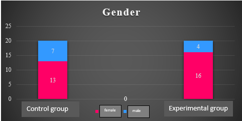
Initially, epidemiological features were analysed. The average age of the control sample was 56.8 (±13.391), minimum 39 years old and maximum 78 years old, and the average age to the experimental sample was 60.55 (±12.713), minimum age being 42 years and maximum 79 years old. As for the gender of patients, both samples were preponderantly female. Thus, female patients in the control sample were in a 65% ratio, and males were 35%, and in the female patients in the experimental sample were in an 80% ratio, and 20% were males (figure 1).
Thereafter, the functional gains achieved by each individual patient, the mean gains for each patient sample and comparatively between the two samples were analysed.
Analysis of data obtained at the initial and final assessments revealed that improvements were seen in both the control sample, and the experimental one, in the sagittal plane cervical spine motion, on both movement directions, flexion and extension, respectively. Tables 1 and figure 2 show the mean gains achieved in the flexion and extension movements between the initial and final tests by the two research samples.
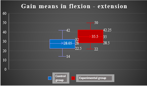
Using the paired-T test for two independent samples to test the differences between the two groups: control and experimental, with regard to the cervical spine movement in sagittal plane, statistically significant differences were obtained (p<0,05), a higher gain mean being noticed in the experimental group (table 2).
Evaluation of data obtained in the initial and final evaluations showed that improvements in the cervical spine movement in frontal plane, on both movement directions, right lateral flexion - left lateral flexion respectively, were reported by both study samples (control and experimental). Table 3 and figure 3 show gain means achieved for the right lateral flexion and left lateral flexion movements between the initial and final tests of the two research groups.
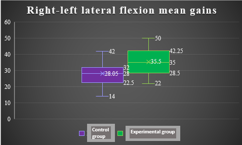
The application of the T-test for two independent samples to test differences between the two groups: control and experimental, with regard to cervical spine movement in frontal plane, revealed statistically significant differences (p<0,05), a higher gain mean being reported by the experimental group (table 4).
Data research from the initial and final evaluations in terms of cervical spine movements in a transverse plane showed that both patient groups registered improvements in the cervical spine movement in this plane, on both movement directions: right- left rotation respectively. Table 5 and figure 4 render the gains mean achieved for right and left rotation movements in between initial and final tests to the two study groups.
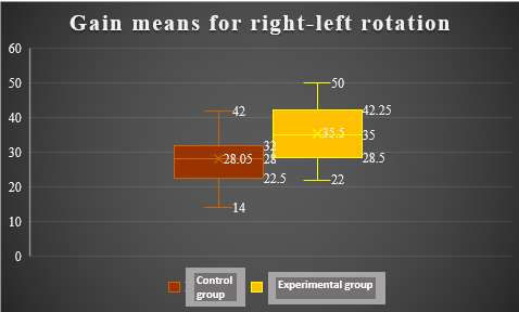
The application of the T-test for two independent samples to test differences between the two study groups, i.e.: control and experimental, with regard to the cervical spine movement in a transverse plane, led to statistically significant differences (p<0,05); however, the experimental group achieved a higher gain mean (table 6).
As for how pain changed or not among the two study groups, data analysis from the two evaluations (initial and final) revealed that it significantly improved in both groups. Also, there were statistically significant differences (p<0,05) in this parameter as well, reported when differences between the two samples were tested using the T-test; however, the experimental group achieved a higher gain mean.
Conclusions
Application of proprioceptive neuromuscular facilitation techniques, in addition to a classic kinetic program, to patients who have cervical osteoarthritis is more effective than just applying a classic kinetic treatment for a term of 24 weeks, respectively: 3 sessions per week in the first 12 weeks and 2 sessions per week over the following 12 weeks.
Introduction of proprioceptive neuromuscular facilitation techniques to the recovery program resulted in increased muscle strength, improved stability and higher range of motion for the cervical spine joints.
Use of a combination of dynamic kinetic techniques and proprioceptive neuromuscular facilitation (slow reversal, slow reversal with opposition, agonistic reversal, rhythmic initiation, rhythmic stabilization, hold-relax exercises) and the manner of organizing the recovery program led to a superior recovery in patients with cervical osteoarthritis from the experimental group.
Cervical osteoarthritis, one of the more and more frequent diagnoses both in the elderly patients, and in the younger ones, requires proper and correctly administered treatment, involving medication to fight off pain and kinesiotherapy to improve mobility and joint stability, but also to reduce pain. By adapting the kinetic program to the patient’s clinical- functional status, improved mobility and stability was achieved, as well as pain reduction, which in turn, resulted in the reduction of medication administration, which has numerous side effects (gastrointestinal, liver, renal and cardiovascular toxicity), especially among the elderly population.
Cervical osteoarthritis presents a symptomatology (especially joint stiffness) impairing the patients’ ability to carry out their daily activities, as this condition progresses into more severe stages. Improvement of the cervical spine’s mobility angles achieved by this study heightened the patients’ functional possibilities, ensuring they are able to carry out their daily physical and recreational activities equally.
References
Al-Youzbaki, D. B., Khalil, N. S., & Tawfeeq, R. S. (2021). Risk Factors of Cervical Spines Osteoarthritis in Adult Women: Case-Control Study. Systematic Reviews in Pharmacy, 12(1), 272-275.
Binder, A. I. (2007). Cervical Spondylosis and Neck Pain. BMJ (Clinical research ed.), 334(7592), 527–531. DOI:
Burcea, C. C., & Ferechide, D. (2013). Methods of data analzsis and processing to allow evincing the knee functional advantage following articular associated lesions related to professional sports activity. Metalurgia International, 18(Special Issue 3), 62-65
Burcea, C. C., Cernușcă-Mitariu, M., Mițariu, M., & Ferechide, D. (2013). Aspects regarding the lower limb recovery and functionality following trochanteric fractures depending on the used implants and biomaterials. Metalurgia International, 18(Special Issue 3), 58-61
Burcea, C. C., Costea, R., Măru, N., Perieanu, V. Ș., Bucur, M. V., Burlibașa, M., Milicescu, Ș., Perieanu, M. V., Malița, M. A., Beuran, I. A., Babiuc, I., Marcov, N., Marcov, E. C., & Drafta, S. (2021a). Applications of Kinetotherapy in the Prophylaxis of Occupational Cervical Syndrome (Overloading) among Professions with a Medical Profile in The Field of Dentistry - Preliminary Study. Acta Medica Transilvanica, 26(4), 72-78. DOI:
Burcea, C. C., Perieanu, M. V., Burlibaşa, M., Costea, R., Perieanu, V. Ș., Tănase, G., Ionescu, C., Babiuc, I., Neagoe, I. C., Costea, R., Mihai, A., Dina, M. N., Maliţa, M. A., Tănase, G., Marcov, E. C., & Chirilă, M. (2020a). The “Sitting” Position and Its Implications in Dental Technology. General Aspects of Improving the Quality of Life - Part II. Acta Medica Transilvanica, 25(4), 66-69. DOI:
Burcea, C. C., Perieanu, M. V., Perieanu, V. Ș., Burlibaşa, M., Tănase, G., Ionescu, C., Babiuc, I., Cristache, C. M., Costea, R., Donciu, I., Oancea, L., Ionescu, I., Marcov, E. C., Maliţa, M., & Beuran, I. A. (2020b). The “Sitting” Position and Its Implications in Dental Technology. General Aspects of Improving the Quality of Life - Part I. Acta Medica Transilvanica, 25(3), 69-74. DOI:
Burcea, C. C., Perieanu, V. Ș., Costea, R. C., Burlibaşa, M., Chirilă, M., Marcov, N., Tănase, G., Măru, N., Farcaşiu, A. T., Milicescu, Ș., Perieanu, M. V., David, M., Ionescu, C., Marcov, E. C., & Moraru, L. (2021b). Applications of Kinetotherapy in the Prophylaxis of Sedentary Behavior among Professions with a Medical Profile in the Field of Dentistry. Acta Medica Transilvanica, 26(2), 52-58. DOI:
de Zoete, R. M., Armfield, N. R., McAuley, J. H., Chen, K., & Sterling, M. (2020). Comparative effectiveness of physical exercise interventions for chronic non-specific neck pain: a systematic review with network meta-analysis of 40 randomised controlled trials. British journal of sports medicine, 55(13), bjsports-2020-102664. Advance online publication. DOI:
Dou, Y., Sun, X., Ma, X., Zhao, X., & Yang, Q. (2021). Intervertebral Disk Degeneration: The Microenvironment and Tissue Engineering Strategies. Frontiers in bioengineering and biotechnology, 9, 592118. DOI:
Ferechide, D., & Burcea, C. C. (2013). Optimisation of physiological recovery in patients with diaphyseal pseudarthrosis of the femur and tibia from the standpoint of identifying the types of metallic osteosynthesis and the biomaterials used in the treatment of these disorders. Metalurgia International, 18(Special Issue 3), 116-118
Ferrara, L. A. (2012). The Biomechanics of Cervical Spondylosis. Advances in orthopaedics, 2012, 493605. DOI:
Kuć, J., & Żendzian-Piotrowska, M. (2020). A Pilot Study Evaluating the Prevalence of Cervical Spine Dysfunction Among Students of Dentistry at the Medical University. Frontiers in neurology, 11, 200. DOI:
Kuo, D. T., & Tadi, P. (2022). Cervical Spondylosis. In StatPearls. StatPearls Publishing. https://www.ncbi.nlm.nih.gov/books/NBK551557/
Louw, S., Makwela, S., Manas, L., Meyer, L., Terblanche, D., & Brink, Y. (2017). Effectiveness of exercise in office workers with neck pain: A systematic review and meta-analysis. The South African journal of physiotherapy, 73(1), 392. DOI:
Lv, Y., Tian, W., Chen, D., Liu, Y., Wang, L., & Duan, F. (2018). The prevalence and associated factors of symptomatic cervical Spondylosis in Chinese adults: a community-based cross-sectional study. BMC musculoskeletal disorders, 19(325). DOI:
Magnus, W., Viswanath, O., Viswanathan, V. K., & Mesfin, F. B. (2022). Cervical Radiculopathy. In StatPearls. StatPearls Publishing. https://www.ncbi.nlm.nih.gov/books/NBK441828/
Saini, A., & Mukhdomi, T. (2022). Cervical Discogenic Syndrome. In StatPearls. StatPearls Publishing. https://www.ncbi.nlm.nih.gov/books/NBK555960/
Singh, S., Kumar, D., & Kumar, S. (2014). Risk factors in cervical spondylosis. Journal of clinical orthopaedics and trauma, 5(4), 221–226. DOI:
Xu, X., Wang, Y., Yang, C., Song, X., Chen, Z., Yang, L., & Li, Y. (2020). Evaluation of rehabilitation effect of five-step exercises on patients with radiculopathy of cervical vertebra. Medicine, 99(26), e20846. DOI:
Copyright information

This work is licensed under a Creative Commons Attribution-NonCommercial-NoDerivatives 4.0 International License.
About this article
Publication Date
10 April 2023
Article Doi
eBook ISBN
978-1-80296-961-0
Publisher
European Publisher
Volume
5
Print ISBN (optional)
-
Edition Number
1st Edition
Pages
1-1463
Subjects
Education sciences, teacher education, curriculum development, educational policies and management
Cite this article as:
Burcea, C., & Cristina Neagoe, I. (2023). New Methodological Approaches In The Rehabilitation Of Patients With Cervical Osteoarthritis. In E. Soare, & C. Langa (Eds.), Education Facing Contemporary World Issues - EDU WORLD 2022, vol 5. European Proceedings of Educational Sciences (pp. 867-875). European Publisher. https://doi.org/10.15405/epes.23045.86