
An Innovative Protocol Of Physiokinetotherapy For The Treatment Of Temporomandibular Disorder

Abstract
Temporomandibular disorder is a very common pathology nowadays and a lot of patients present this specific symptomatology. Because of its plurifactorial cause, the treatment has to be a complex one that includes oral rehabilitation of the occlusion and physiokinetotherapy of the temporomandibular joint. In association to the treatment, for better results, the international literature mentions the benefits of the laser. The treatment protocol begins with impressions for models, then determining the cranial maxillary position by using the face bow, mounting the models in order to analyse the case to determine the correct centric relationship of the patient. Personalised splints are fabricated with the models in the SAM 2 articulator, the role of the casts is to maintain the mandibular condyle in the most anterior superior position in the glenoid cavity. The next step in the protocol begins with the application of the splints, readjusting the occlusal contacts at the level of the splint at five days after the application of the splint, then every week, associated with kinetotherapy exercises, opening-closing movements, protrusion, laterality and movements for the scapular-cervical girdle in order to regain the articular mobility and to be able to open the mouth. Together with the exercises, we need laser biostimulation at the level of the temporomandibular joint in order to decrease inflammation, pain and to induce muscle relaxation. By doing all these procedures, we will have a better understanding of the occlusal factors that have produced changes in the temporomandibular joint.
Keywords: Physiokinetotherapylaser biostimulationsplint
Introduction
The temporomandibular joint represents a synovial type of joint that consists of the condyle of the mandible, the articular eminence of the temporal bone and the articular disc that divides the articular space in two cavities: a superior one and an inferior one (Rasavong, 2008).
The role of the articular disc is to prevent the harm of the articulating surfaces, to absorb and after that to scatter the load effect on a wider area of contact. This fact has a very big significance in order to preserve the health of the stomatognathic system and also to avoid the alterations of the articular disc that may lead to the appearance of temporomandibular disorders (TMDs). (Ferreira, Simamoto-Júnior, Soares, Ramos, & Fernandes-Neto, 2017)
The TMDs are as well called craniomandibular disorders and comprise a class of pathologies that alter the masticatory muscles, the temporomandibular joint and also the structures related to it. This type of disorders affects a percentage of more than 25% of the general population (McNeely, Armijo Olivo, & Magee, 2006; McNeill, 1993; Di Fabio, 1998; Gremillion, 2000).
The most common signs and symptoms of the TMDs are pain in the joint area, joint sounds, limitation in different jaw movements and functions, muscle and joint tenderness, catching/locking of the jaws and it can also be correlated with symptoms in the head and neck region that include headache, facial pain, cervical spine disorders and symptoms in the ear region (Benoit, 1994; Chen, Liu, Yap, & Fu, 2017).
The temporomandibular functional joint disorders can be ranked as derangements of the condyle-disc complex, structural incompatibility of the articular surfaces, which can be classified as disc-interference disorders and inflammatory joint disorders (Okeson, Perez, & Fricton, 2017).
There are four types of articular disc displacements, according to the Diagnostic Criteria for Temporomandibular Disorders: disc displacement with reduction, disc displacement with reduction with intermittent locking, disc displacement without reduction with limited opening and disc displacement without reduction without limited opening (Pihut, Gorecka, Ceranowicz, & Wieckiewicz, 2018).
Because of its plurifactorial cause, the treatment of TMD has to be a complex one that involves a multidisciplinary approach that includes the collaboration of different specialists: dentists, orthodontists, psychologists, physical therapists, physicians and neurologists. Physical therapy was cited to be a significant treatment by the American Academy of Craniomandibular Disorders and the Minnesota Dental Association (McNeely, Armijo Olivo, & Magee, 2006).
For better results of the therapy, it can be associated with anterior repositioning splints/occlusal splints and laser biostimulation.
Together with the exercises and the splints, we need laser biostimulation at the level of the temporomandibular joint in order to decrease inflammation, pain and to induce muscle relaxation.
The role of physical therapy is to calm musculoskeletal pain, diminish inflammation and improve the oral motor function. The interventions used for the treatment of TMDs include electrophysical modalities such as ultrasounds, microwave, laser, transcutaneous electrical nerve stimulation, exercises and manual therapy techniques. (McNeely, Armijo Olivo, & Magee, 2006)
The main role of exercises is to improve coordination of the muscles involved in the mastication process, diminish the muscle spasm and modify the pattern of the jaw closure (Nicolakis et al., 2001).
The most important fact is to understand the aetiological factors that determine the TMDs, which include trauma, parafunction, arthritis and generalised muscle hyperactivity. The initial therapy of these pathologies lies in the reduction of the jaw use by adopting a soft diet and the restraint of the jaw movement to within the pain-free range, the use of non-steroidal anti-inflammatory drugs and self- utilisation of heat or ice packs, as required in order to obtain certain relief. The secondary therapy consists in the behaviour modification, which trains the patient to avoid the painful habits of tooth clenching. (Clark, Seligman, Solberg, & Pullinger, 1990)
The use of splints is thought-out as a reversible non surgical treatment modality because of its property to diminish pain at the level of the temporomandibular joint, pain that is induced by too much occlusal pressure determined by external forces. The use of splints improves the blood circulation at the level of the temporomandibular joint and preserves a wide gap between the mandibular condyle and the mandibular fossa. (Hegab, Youssef, Al Hameed, & Karam, 2018).
Problem Statement
Temporomandibular disorder is a very common pathology nowadays, a lot of patients presenting this specific symptomatology that influences their quality of life.
Research Questions
We want to check an innovative protocol of physiokinetotherapy for the treatment of temporomandibular disorder. The proposed protocol is non invasive, has low cost and acts on multiple factors implicated in the aetiology of the temporomandibular disorder. The innovation of the proposed protocol lies in combining three therapeutic instruments accepted worldwide as efficient in the treatment of TMD. Personalised splints position the mandibular condyle in the most antero-superior position and shortcut the determined dental position. Laser therapy acts on muscle relaxation and, together with exercises, can lead to obtaining a new correct position of the mandible.
Purpose of the Study
The aim of this study was to present an innovative protocol that included manual therapy, exercises, occlusal splint and laser biostimulation for the treatment of a bilateral disc displacement with reduction using a single-case design.
Research Methods
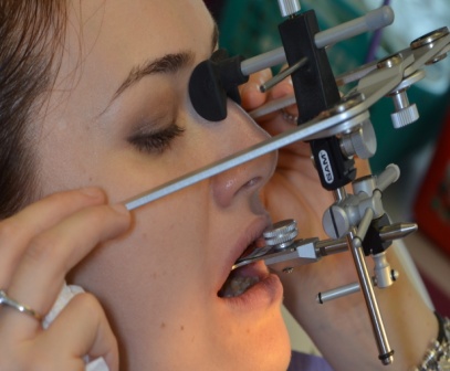
The subject of our research was a 34-year-old female patient that was referred to our department for dental mobility of the tooth 2.1., crackles, abfraction lesions, bruxism with jaw clenching, headache and temporomandibular joint pain. The treatment protocol begins with impressions for models, then it is determined the facial report by using the face bow, mounting the models in order to analyse the case to find the correct centric relationship of the patient, as shown in Figure
The next step in the protocol begins with the application of the splints, readjusting the occlusal contacts at the level of the splint at five days after the application of the splint, then every week. The kinetotherapy consisted of daily exercises, performed twice a day. The patient performed before a mirror for self-correction, exercises such as opening-closing movements, protrusion, laterality and movements for the scapular-cervical girdle.
Findings
The medical and dental histories of the patient were recorded. The written and informed consent was obtained. The patient’s history revealed that the TMJs pain and headache started insidiously one year ago, her symptoms being, at the beginning, at the level of the TMJs than the head. The pain was not exacerbated by the opening of the mouth.
At the moment of the examination, the patient reported constant pain in her temporomandibular joint; she also noted a history of occipital headaches that initiated soon after she felt the jaw pain.
She was not under any medication treatment, but only occasionally she took a non-steroidal anti-inflammatory drug. She described bruxism in her medical history, denying any systemic disease.
Examination of the mouth opening did not increase the patient’s symptoms and she was able to open and close the jaws without any pain. The lateral excursion of the mandible was restricted to 3 mm on the right side and 4 mm on the left side, and the mandibular protrusion was restricted to only 3 mm. The auscultation of the temporomandibular joint revealed joint clenching and also crepitus bilaterally.
Hypomobility of the C6-T2 segment and also the C1-C3 increased the symptoms in the occipital area of the patient.
At the extra oral examination, we found bilateral tenderness in the masseter muscle, restrictions during the mandibular depression in translation at the condyle level.
Palpation of the trigeminal and cranial nerves revealed no alteration of their function or sensibility of the innervated region.
The treatment protocol begins with impressions for models (taken with impression material – Alginat), followed by the determination of the cranial maxillary report by using the face bow, which is needed for semi-adjustable articulators. In order for the maxillary and mandibular models to be mounted in a correct relation to the mandibular joints, the face bow is used.
Then, the models were mounted in a semi-adjustable articulator in order to design the splints in the most superior and anterior positions of the mandibular condyle.
The next step in the protocol begins with the application of the splints, readjusting the occlusal contacts at the level of the splint at five days after the application of the splint, then every week, for a total of three months, associated with kinetotherapy exercises, opening-closing movements, protrusion, laterality and movements for the scapular-cervical girdle in order to regain the articular mobility and to be able to open the mouth.
The treatment phase consisted of 10 visits, one per week; during this period, we also performed 5 sessions, one per week, of laser biostimulation with the Fotona Light Walker laser (Ljubljana, Slovenia), Fotona Genova Hand piece, mode MSP (micro short pulse), 0.5 W, 10 Hz, for 60 seconds per application point. We performed only five sessions, because the symptomatology decreased at the level of both the jaws and the head. On a Visual Analogue Scale (VAS), the initial pain measured in the first session was 8, after 3 sessions it decreased to 4, and at the end of all treatment sessions, there was no pain.
Exercises regarding the postural education of the patient by using manual physical techniques aimed at the rehabilitation of the temporomandibular joint and the scapular-cervical girdle.
The treatment protocol included anterior glides and distractions of the bilateral temporomandibular joint. Another exercise, namely a jaw opening exercise, included the placement of all fingers (without the thumb) of the dominant hand on the incisal margin of the mandibular teeth, while the other fingers were placed on the maxillary incisal margin of the frontal teeth. The mouth was opened this way, while the patient felt the masticatory muscle tightness, and that position was held for 10 seconds.
Exercises for the neck included rotation of the head to the right side and set with the same hand at the level of the left ear, and then pressing against the head position with the right hand and extension of the neck until the tightness of the muscle was felt; this position was kept for 10 seconds too. The patient also performed isometric cervical flexion forward. (Cleland & Palmer, 2004; Ishiyama et al., 2017)
The exercises performed by the patient led to muscular relaxation of the cervicofacial muscles, allowing for better mobility, reducing pain and increasing patients’ comfort and quality of life.
Conclusion
By applying this protocol to our patient, improvement was observed in the symptoms and clinical signs, quantified in pain relief, mobility of the head and neck and also the tooth 2.1, the muscle contraction and tenderness were reduced, even the physiology and contour of the patient’s face improved.
The mandible was positioned anteriorly because of the use of the splint, correcting the occlusion, and at six month after the use of the splint, a new determination made possible the correct occlusion by eliminating the interference and premature contacts.
This protocol is applicable to most patients with TMJ problem, but a very accurate diagnosis has to be established before entering this protocol treatment. More clinical evidence has to be brought, but this is a first step in our research.
References
- Benoit, P. (1994). History and physical examination for TMD. In S. L. Kraus (Ed.), Clinics in Physical therapy: Temporomandibular disorders (2nd ed.) (pp. 71-98). New York, NY: Churchill Livingstone.
- Chen, H. M., Liu, M. Q., Yap, A. U. J., & Fu, K. Y. (2017). Physiological effects of anterior repositioning splint on temporomandibular joint disc displacement: A quantitative analysis. Journal of Oral Rehabilitation, 44(9), 664-672.
- Clark, G. T., Seligman, D. A., Solberg, W. K., & Pullinger, A. G. (1990). Guidelines for the treatment of temporomandibular disorders. Journal of Craniomandibular Disorders, 4(2), 80-88.
- Cleland, J., & Palmer, J. (2004). Effectiveness of manual physical therapy, therapeutic exercise, and patient education on bilateral disc displacement without reduction of the temporomandibular joint: A single-case design. Journal of Orthopaedic & Sports Physical Therapy, 34(9), 535-548.
- Di Fabio, R. P. (1998). Physical therapy for patients with TMD: A descriptive study of treatment, disability and health status. Journal of Orofacial Pain, 12(2), 24-35.
- Ferreira, F. M., Simamoto-Júnior, P. C., Soares, C. J., Ramos, A. M. A. M., & Fernandes-Neto, A. J. (2017). Effect of occlusal splints on the stress distribution on the temporomandibular joint disc. Brazilian Dental Journal, 28(3), 324-329.
- Gremillion, H. A. (2000). The prevalence and etiology of temporomandibular disorders and orofacial pain. Texas Dental Journal, 117(7), 30-39.
- Hegab, A. F., Youssef, A. H., Al Hameed, H. I. A. A., & Karam, K. S. (2018). MRI-based determination of occlusal splint thickness for temporomandibular joint disk derangement: A randomized controlled clinical trial. Oral Surgery, Oral Medicine, Oral Pathology and Oral Radiology, 125(1), 74-87.
- Ishiyama, H., Inukai, S., Nishiyama, A., Hideshima, M., Nakamura, S., Tamaoka, M., & Wakabayashi, N. (2017). Effect of jaw-opening exercise on prevention of temporomandibular disorders pain associated with oral appliance therapy in obstructive sleep apnea patients: A randomized, double-blind, placebo-controlled trial. Journal of Prosthodontic Research, 61(3), 259-267.
- McNeely, M. L., Armijo Olivo, S., & Magee, D. J. (2006). A systematic review of the effectiveness of physical therapy interventions for temporomandibular disorders. Physical Therapy, 86(5), 710-725.
- McNeill, C. (1993). Temporomandibular disorders: Guidelines for classification, assessment and management (2nd ed.) (pp. 19-22). Chicago, Ill: Quintessence Publishing Co.
- Nicolakis, P., Erdogmus, B., Kopf, A., Ebenbichler, G., Kollmitzer, J., Piehslinger, E., & Fialka‐Moser, V. (2001). Effectiveness of exercise therapy in patients with internal derangement of the temporomandibular joint. Journal of Oral Rehabilitation, 28(12), 1158-1164.
- Okeson, J. P., Perez, C., & Fricton, J. R. (2017). Temporomandibular joint disorders. In J. N. A. R. Ferreira, J. Fricton & N. Rhodus (Eds.), Orofacial disorders: Current therapies in orofacial pain and oral medicine (pp. 145-157). Springer, Cham.
- Pihut, M., Gorecka, M., Ceranowicz, P., & Wieckiewicz, M. (2018). The efficiency of anterior repositioning splints in the management of pain related to temporomandibular joint disc displacement with reduction. Pain Research and Management, 2, 1-6.
- Rasavong, C. (2008). Temporomandibular joint exercise prescription for physical therapists. Retrieved from http://www.cyberpt.com/tmj.asp
Copyright information

This work is licensed under a Creative Commons Attribution-NonCommercial-NoDerivatives 4.0 International License.
About this article
Publication Date
16 February 2019
Article Doi
eBook ISBN
978-1-80296-054-9
Publisher
Future Academy
Volume
55
Print ISBN (optional)
-
Edition Number
1st Edition
Pages
1-752
Subjects
Sports, sport science, physical education
Cite this article as:
Bordea, R., Lucaciu, O., Sîrbu, A., Crișan, B., Bran, S., & Câmpian, R. S. (2019). An Innovative Protocol Of Physiokinetotherapy For The Treatment Of Temporomandibular Disorder. In V. Grigore, M. Stănescu, M. Stoicescu, & L. Popescu (Eds.), Education and Sports Science in the 21st Century, vol 55. European Proceedings of Social and Behavioural Sciences (pp. 516-521). Future Academy. https://doi.org/10.15405/epsbs.2019.02.64