
Mitotic Epithelial Of The Lens Of Rainbow Trout Exposed To Benzene Compounds


Abstract
The effect of 1,3,5 -trichlorobenzene, nitro-benzene, and 2-naphthol on the mitotic activity and frequency of micronuclei in the differentiated zone of the lens epithelium of the rainbow trout Oncorhynchus mykiss Rich was studied in chronic Richopiates. It was shown that the number of micronuclei in the preequatorial zone of the crustal epithelium associated with the properties of the toxicant to influence the proliferative activity of cells in the vegetative zone. It was found that low concentrations of nitrobenzene and 2-naphthol act as inhibitors of mitotic activity and lead to a decrease in the frequency of occurrence of micronuclei in the differentiated zone of the lens epithelium. As the concentration increases, all the studied benzene compounds dissolved in an aqueous medium stimulate mitotic activity, and the number of micronuclei increases. It is concluded that benzene compounds do not behave as radiomimetic and do not cause chromosome breakage during mitosis. The increase in the number of micronuclei, which depends on the number of chromosomal aberrations, occurs due to random errors, the number of which increases, and stimulation of mitotic activity. Consequently, the micronucleus test (MCT) reveals spontaneous aberrations of chromosomes, the number of which increases with the stimulation of cell proliferation in the lens epithelium. Based on this, many substances that were previously classified as genotoxicants because of the MNT are actually stimulators of mitotic activity, and the micronucleus test for most substances, except for radiomimetic, should be considered not cytogenetic, but cytotoxic.
Keywords: Benzene compounds, Genotoxicity, lens epithelium, micronuclei, mitotic activity, Oncorhynchus mykiss
Introduction
Benzene compounds are now increasingly entering commercial water bodies with industrial wastewater. This is facilitated by both the expansion of production of chlorobenzene and nitrobenzene compounds in the chemical industry, and the low susceptibility of nitrobenzene and chlorobenzene compounds to biological destruction in wastewater treatment plants (Bespamyatnov et al., 1975; Dolivo-Dobrovolsky, 2016).
Lower vertebrates are less resistant to chlorobenzene and nitrobenzene compounds than birds and mammals (Oettingen, 1941). In addition, benzene compounds, when ingested in separate portions, disrupt intimate cellular processes (Palade & Goldstein, 1960). One of these processes can be considered mitosis, in which complex molecular and sub molecular rearrangements occur in genetic material and especially in chromosomes, leading to spontaneous aberrations of chromosomes, their fragmentation and lag. Once these fragments and abnormal chromosomes are wrapped in a shell, they turn into microkernels.
The long-term effect of low doses of toxic substances on mitotic activity and cytodifferentiation in hydrobionts significantly differs from the effect of high concentrations of harmful substances, which are most often studied in toxicological laboratories. Under the action of high concentrations of the toxicant, mitotic activity, can generally be suppressed, and in this case, due to the absence of mitoses, micronuclei may not form. Under the influence of low concentrations of harmful substances, the body includes reparative systems that allow it to maintain the entire living system in a relatively satisfactory state in physiological terms. Most often, reparative processes are accompanied by stimulation of mitotic activity. In this case, an increase in the frequency of occurrence of micronuclei in the cells of the studied tissue can give the toxicologist very useful information, since an increase in the number of micronuclei is associated with mitotic activity, and indicates genome instability. For the toxicologist, it is also important that the change in the frequency of occurrence of micronuclei is a quantitative indicator, using which it is possible to give a more objective assessment of the degree of impact of harmful substances on the genetic apparatus of the studied organisms.
In addition, the task was also to study the possibility of using the fish lens in chronic toxicological experiments, which makes it possible to determine the permissible concentrations of harmful substances when establishing fisheries management criteria. MPC for such an important biological indicator as cytotoxicity. This is facilitated by the fact that the micronuclei formed during mitosis in the germinal zone of the lens epithelium accumulate in differentiated cells and are closed for a long time, before the epithelial cells turn into lens fibers.
Problem Statement
The fish lens can be considered one of the most convenient systems for studying the effect of small doses of toxicants on genome stability and cytodifferentiation. This is a pure population of cells, similar to a culture where nerves and blood vessels are absent. At the same time, this system is highly ordered, which is not typical of any artificially maintained human cell culture. Mitoses in the lens epithelium of fish are observed throughout life, and in young organisms mitotic activity is more pronounced.
Purpose of the Study
We can expect an increased formation of micronuclei in the lens epithelial cells of juvenile fish, sin micronuclei are formed during cell division. For this reason, we set out to conduct studies on fingerlings of the rainbow trout Oncorhynchus mykiss Rich., which allowed us not only to establish the effect of small doses of toxicants on the frequency of micronucleus formation in lens epithelial cells, but also to compare this effect in different areas of epithelial differentiation.
Research Methods
The study was conducted on rainbow trout cutlets weighing 1-3 g and measuring 9.5 cm in length. Fish were placed in 1.55-liter aquariums of 5 specimens each. The water temperature in aquarium was maintained in the range of 16-170 C (Nikiforov‐Nikishin, Nikiforov‐Nikishin, et al., 2021). The water was continuously aerated and changed every other day. The trout heads were fed using French Esturgeon feed». The experiment duration was 21 days. For the experiment, 18 aquariums were used, of which three served as control ones, the fish lived in sewage tap water, and in the remaining aquariums the fish were kept in solutions of benzene compounds. For each compound, 5 concentrations were taken, the spectrum of which was determined depending on the established sanitary and hygienic standard for the content of harmful substances in the water of MPC. These concentrations are higher than the established for the same substances. However, they proved to be effective for trout fingerlings, which makes it possible to identify the relationship between mitotic activity in different areas of lens epithelial cytodifferentiation and the frequency of micronuclei occurrence.
The effect of such compounds as1,3,5 -trpchlorobenzol, 2-naphthol, and nitrobenzene on the mitotic activity of various zones of the lens epithelium was studied in a chronic experiment. All these compounds affect cell proliferation, and 2-naphtholalso has mutagenic properties. Five concentrations were taken for each compound, and one of the concentrations corresponded to the established sanitary and hygienic MPCB (Bespamyatnov et al., 1975). Concentrations of 0.1, 0.07, 0.05, and 0.03 were taken for tri-chlorobenzene: 0.01 mg/l. MPCB= 0.03 mg/l. For 2-naphthol, the concentrations are taken 0,8; 0,6; 0,4; 0,2; 0,1 MPCB = 0.4 mg / l. Nitrobenzene was added to aquariums at the same concentrations as 2-naphthol. The maximum permissible-nitrobenzene is 0.2 mg/l.
If the maximum permissible concentrations of the studied benzene compounds are lower. If the maximum permissible concentration (MPCB) is determined, then this will indicate a high sensitivity of the proposed method, and it can be recommended for establishing permissible concentrations of pollutants for cytotoxic indicators when establishing a fisheries standard for MPCpx. ОIn general, theMPC rx standardрх is more strict, and 10-15 times lower, than the MPCb.
After the experiment, the fish were slaughtered, and their eyes were fixed in the mixture Carnois. The eyes of the control fish were recorded simultaneously before the experiment and at the end of the experiment. Lenses were removed from fixed eyes and squamous preparations were prepared from their epithelium for cytological studies (Howard, 1952). Mayer's gemalown was also used for staining the preparations. The mitotic index (MI) was calculated in the lens epithelium) and the number of micronuclei in the germinal and preequatorial zones. For this purpose, six fields bounded by the ocular mesh were calculated on each preparation (three fields in the germinal zone and three fields in the preequatorial zone). In the differentiated zone, the cells were arranged in rows for further ordered transformation into lens fibers. The layout of the calculated fields in the lens epithelium is shown in Figure 1.
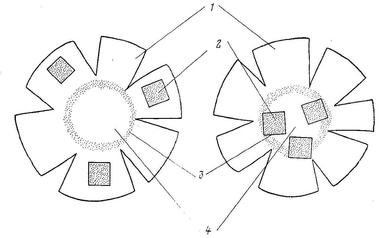
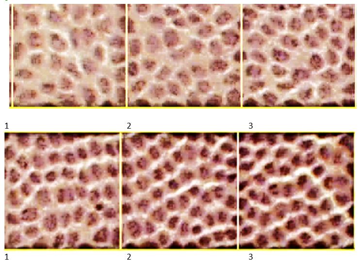
On each preparation, the mitotic index (MI) was calculated in the germinal zone of the epithelium in three fields, isolated by the ocular mesh in 100 cells. Taking into account the presence of ten total epithelial preparations from five fish, the total cell count for detecting MI and micronuclei in the germinal zone of the epithelium for each concentration was about 1000. Accordingly, MI and the frequency of occurrence of micronuclei in the preequatorial zone were determined when counting 1500 cells, since their density was higher in the same area. An oculi was used to accurately localize a particular zone of differentiation of the monolayer epithelium of the lens bright grid with a division price of 0.1 mm, so that one square in the germinal zone included 33-35 cells, and in the pre-equatorial zone 47-50 cells, with an increase of 12.5 x 40. The frequency of occurrence of micronuclei and mitoses in a certain area of cytodifferentiation of the lens epithelium was calculated in%. The first three zeros captured poorly differentiated areas of the epithelium, in which cell division occurred (square 2), and spatial rearrangement of cells, for example, the formation of ring structures, as in square 3 (Figure 2). Figure 2 (top) shows a band of cells, extending from the center to, the periphery of the lens epithelium, bounded by the ocular cross-point, which includes part of the primary zone (1), the germinative zone (2), and the zone of ring structures (3). At the bottom, three sections of the preequatorial zone are shown, where cells are arranged in rows and prepared to form lens fibers. The MI and frequency of occurrence of micronuclei were determined in squares 2 and 5.
Based on the results obtained, the reliability of the difference in mean MI for different concentrations of toxic compounds was calculated. Statistical processing for low-and high-differentiated zones was carried out according to the Student's criterion.
Findings
In the control preparations of the lens epithelium of rainbow trout fingerlings in the germinal zone, the mitotic index (MI) it is equal to 5.97, and the frequency of occurrence of microkernels is 4.3. In the preequatorial zone of the lens epithelium, where cell differentiation is advanced towards the formation of lens fibers, MI drops sharply to 1.53. At the same time, the number of microkernels reaches 14.4%, as they accumulate and persist in zone 5 (Figure 2). Cells with pycnotic nuclei are also found in the highly differentiated zone. No changes in the mitotic index were detected in the control fish during the chronic experiment (21 days).
Under the action of trichlorobenzene, 2-naphthol, and nitrobenzene, changes in the mitotic index and frequency of micronuclei were observed to decrease or increase, depending on the substances and their concentration.
Under the influence of trichlorobenzene ash in the lowest concentrations (0.01 and 0.03 mg/l), the mitotic activity in the lenses practically does not differ from the control. At the same time, within the limits of statistical unreliability, the frequency of occurrence of micronuclei in the germinal and pre-equatorial zones fluctuates. Consequently, low concentrations of trichlorobenzene do not significantly affect the mitotic index (MI) and the frequency of occurrence of micronuclei in the lens epithelium. Only at a trichlorobenzene concentration of 0.05 mg/l does a significant difference appear between the control and the experiment. At the same time, MI in the genezone rises to 9.2.44, and the frequency of occurrence of micronuclei reaches 6.4.
When the trichlorobenzene concentration increases to 0.07 mg/L, the mitotic activity in the germinal zone increases almost twofold compared to the control (MI =11.8), and at the same time the frequency of occurrence of microkernels increases. In the pre-equator zone, MI also increases and becomes slightly higher than the control value. However, in this differentiated zone of the lens epithelium, the frequency of occurrence of micronuclei increases and reaches 25,625.6%. However, this increase occurs less than twice, as it is noted in the germinal zone. The highest mitotic activity under the influence of trichlorobenzene at a concentration of 0.1 mg / l is observed in poorly differentiated cells of the lens epithelium, in the germinal zone. At the same time, the number of micronuclei in the preequatorial zone increases (Table 1).
The effect of 2-naphthol has a twofold effect on the mitotic activity and frequency of occurrence of micronuclei in the lens epithelium. Low concentrations also have an inhibitory effect on mitotic activity and a decrease in the number of micronuclei in the germinal zone. At the same time, in the highly differentiated preequatorial zone, where only rare mitoses occur. the number of mitoses dropped about even more. At the same time, the number of micronuclei also decreased compared to the control. Apparently, these preserved microkernels were formed even before the experiment was set up.
и2-naphthol concentrations are also high (0.6 and 0.8 mg / l) how would the inhibitory effect of the studied pollutant be removed? At a concentration of 0.6 mg/l. in the germinative zone, as small amount of MI is observed up to 7.10, and the frequency of occurrence of bl micronuclei is close to control. In the preequatorial zone of the epithelium, mitotic activity does not even reach the control parameters, and often the occurrence of micronuclei is close to the control.
Under the influence of a 2 - naphthol concentration of 0.8 mg/l in the germinal zone, cell proliferation is stimulated (MI= 10.4). The number of micronuclei also increases statistically significantly in relation to the control. In the preequatorial zone, the frequency of occurrence of micronuclei increases due Toth accumulation of cells with chromosome abnormalities that came from the germinal zone, but the number of mitoses is comparable to the control (Table 2).
Nitrobenzene only in the lowest concentration studied (0.1 mg/l) has an inhibitory effect on mitotic activity and the amount of micronucleus formation in the germinal and preequatorial zones of the lens epithelium. Starting from concentrations above 0.2 mg / l, only the stimulating effect of the test substance on mitotic activity and micronucleus formation is observed in the germinative and highly differentiated zone. As all the previous compounds studied, nitrobenzene has the greatest effect on the small differentiated zones of the lens epithelium. Data on the effect of nitrobenzene on the mitotic index and the frequency of micronuclei in various zones of differentiation of the fish lens epithelium are presented in Table 3.
The main feature of the results of this study is that: It is shown that the frequency of occurrence of micronuclei in the fish lens epithelium is considered depending on the cytodifferentiation and mitotic index (MI), which changes under the action of benzene compounds. A micronucleus test that was previously performed on fecal, normal epithelium, or blood cells. It does not take into account factors related to cell cytodifferentiation and mitotic activity. The lens epithelium has the advantage that cells with micronuclei are not rejected, but are preserved in the preequatorial zone. Previously developed methods of conducting a micronucleus test allow us to study only the upper layers of differentiated cells of the multilayer epithelium, without taking into account mitotic activity, which could be detected on sections of the multilayer epithelium. But on the slice, we could not collect the necessary number of cells to account for the occurrence of micronuclei. Thus, it becomes possible to represent the single-layer epithelium of the lens as a section of the multilayer epithelium, in which all cells with micronuclei are preserved in a highly differentiated layer. We can assume, that the single-layer epithelium of the lens allows us to look at a section of the multilayer epithelium,
It is shown that the frequency of occurrence of micronuclei depends on mitotic activity in the germinative zone, which is determined by the action of a particular benzene compound. The action of trichlorobenzene ash differs from that of 2-naphthola p nitrobenzene. While trichlorobenzene at low concentrations under study has no effect on micronucleus formation, and with increasing concentrations acts as a stimulator of mitotic activity and the frequency of occurrence of micronuclei, 2-naphthol and nitrobenzene in small concentrations under study inhibit mitotic activity and, accordingly, contributes to a decrease in micronucleus formation. 2-naphthol has even more pronounced inhibitory properties than nitrobenzene.
Analysis of the effects of three benzene compounds shows a clear dependence of the formation of micronuclei on MI and on the cytodifferentiation of cells in a particular area of the lens. At the same time, there is a direct dependence of the formation of micronuclei in epithelial cells on MI. All this forces us to take a different look at the detection of genotoxicity of substances with the newly developed MIN tests, which take into account only the number of micronuclei. Regardless of mitotic activity. With this approach, many substances that stimulate mitotic activity can be perceived as genotoxicants that cause chromosomal aberrations (Nikiforov-Nikishin, Shatokhin, et al., 2019). In fact, chromosomal aberrations, as a result of which the frequency of occurrence of micronuclei increases, they occur indirectly due to the stimulation of mitotic activity and the appearance of a greater number of spontaneous mutations in chromosomes. Most likely. chromosome fragmentation can only be caused by radiolabeled substances or compounds that disrupt the mitotic mechanism. There are significantly fewer such chemical compounds than substances that manifest themselves as stimulators of mitotic activity, including the benzene compounds we studied. The mechanism of the effect of benzene compounds on crustal epithelial cells has not yet been sufficiently studied. Perhaps the capsule, which has selective permeability, plays an important role in the action of substances on the lens epithelium (Leary & Van Geyningen, 1968). It may turn out that the effect of benzene compounds on the mitotic activity of the fish lens epithelium is mediated. It is known that low doses of the toxicant have a stimulating effect, so under the influence of low concentrations of the studied substances, the stimulating effect can also affecting increase in the mitotic index in the lens epithelium.
The opposite assumption is also quite acceptable; according to which mitosis is inhibited by halons [interleukin] (Bullough & Laurence, 1960; Simakov et al., 2020). Later, Bullough (1971) expanded his theory and [the functions of halons included not only the regulation of mitotic activity, but also the regulation of growth and differentiation. Phenolsand aromatic compounds are known to play a major role in chemical carcinogenesis (Glebova et al., 2019; Suess et al., 1977). From this point of view, the studied benzene compounds can be considered as products that exhibit some properties of chemical carcinogens. In particular, under the influence of such compounds, cellular receptors that perceive halons are blocked, and cells begin to divide intensively. Further studies in this direction may make the lens epithelium of fish and other vertebral animals a test object that allows us to judge the cytotoxicity of the test substance, the stimulating effect of the substance should be considered, as a manifestation of cytotoxicity.
Analysis of the obtained results on the effect of benzene compounds on the mitotic activity of the trout lens epithelium shows that the mitosis-stimulating effect begins to manifest itself from tho second centration’s that are close to the sanitary and hygienic MPCB. For trichlorobenzene, mitosis stimulation in the germinative zone begins to increase when concentrations are higher than 0.03 mg/l (at 0.05 mg / L), for 2-naphthol - with 0.5, and for nitrobenzene — with 0.2 mg/l. However, the epithelium of the lens of rainbow trout is not mitotically indifferent to concentrations lower, than the sanitary and hygienic macs. At all lower concentrations, there is a change in the mitotic index in the epithelium and lens. In one case, these changes are insignificant and statistically unreliable compared to the control, as in trichlorobenzene. a2-naphthol and nitrobenzene act as inhibitors of mitotic activity at the lowest concentrationsstudied.2-naphthol has the most pronounced phpb-forming property.
The rate of decrease or increase in the frequency of occurrence of micronuclei in the preequatorial zone of the lens epithelium lags behind the fluctuations of MI in the germinal zone. This can be explained by the fact that, most likely, for 21 days of the experiment, the accumulation of micronuclei in the preequatorial zone depends on the arrival of cells with micronuclei from the germinal zone. Apparently, the number of micronuclei in a more differentiated area of the lens epithelium will increase in the future. We investigated only concentrations close to the threshold and maximum permissible values. These are relatively low concentrations, and when they act, both inhibition and stimulation of mitosis are noted. At higher concentrations, the next drop-in mitotic activity can be expected. Thus, these studies confirm the phasing effect of toxicants at low concentrations - a phenomenon that developers of sanitary and fisheries standards often encounter MPC.
Of the studied substances (for trichlorobenzene 0,001 mg / l, for 2-naphthol 0,05 mg/l, for nitrobenzene 0,01 mg/l). which is about an order са of magnitude lower than the sanitary and hygienic requirements. MPCin. Our research confirms that fish, as a test object, are more sensitive to toxicants than rats, which are subject to sanitary macs. For nitrobenzene and 2-naphthol, concentrations even below MPC be effective. Taking this circumstance into account, the trout lens can be recommended for use as a test object in the system for determining of harmful substances for fish reservoirs
Conclusion
At concentrations of trichlorobenzene 0,05; 0,07; 0,1 mg / l, 2-naphthol0.6 p 0.8 p nitrobenzene 0.4; 0.6; 0.8 mg / L, mitosis stimulation is observed in the germinal zone of the lens epithelium of rainbow trout cells and simultaneously an increase in the frequency of micronuclei in the highly differentiated preequatorial zone.
At lower concentrations of the studied substances, the frequency of occurrence of micronuclei in differentiated cells either does not change (chlorobenzene), or the compounds have a stimulating effect on mitoses and the frequency of occurrence of micronuclei (2-naphthol and nitrobenzene).
The regularity of micronucleus increase in the preequatorial zone of fish lens epithelium with increasing mitotic index in the germinal zone was revealed. This may be due to an increase in the number of dividing cells and an increase in spontaneous chromosomal aberrations.
Given the high sensitivity of the germinal zone to changes in mitotic activity, the trout lens can be recommended for use as a test object in aquatic toxicology, especially for substances that inhibit or stimulate mitoses.
References
Bespamyatnov, G. P., Bogushevskaya, K. K., Bespakhyakova, A. V., Krotov, Yu. A., Zelenskaya, L. A., Plekhotkin, V. F., & Smirnov, G. G. (1975). Maximum permissible¬concentrations of harmful substances in air and water. Khimiya.
Bullough, W. S. (1971). Ageing of mammals. Nature, 229(5287), 608–610. DOI:
Bullough, W. S., & Laurence, E. B. (1960). The control of epidermal mitotic activity in the mouse. Proc. Boy. Soc. (B), 151, 517-536. DOI:
Dolivo-Dobrovolsky, L. B. (2016). Microbiological processes of water purification. Publishing House of the Ministry of Communal Services of the RSFSR.
Glebova, I. A., Nikiforov-Nikishin, D. L., Gorbunov, A. V., Kozlov, A. V., Gybina, T. G., Zaitseva, N. A., & Larionova, A. A. (2019). Investigation of the Presence of Mutagens in the Coastal Part of Lake Senezh near the Highway. Ekoloji, 28(107), 385-90.
Howard, A. (1952). Whole mounts of rabbit lens for cytological studv. Stain technol., 27, 313-317. DOI:
Leary, A., & Van Geyningen, R. (1968). Biochemistry of the eye. Meditsina.
Nikiforov‐Nikishin, A., Nikiforov‐Nikishin, D., Kochetkov, N., Smorodinskaya, S., & Klimov, V. (2021). The influence of probiotics of different microbiological composition on histology of the gastrointestinal tract of juvenile Oncorhynchus mykiss. Microscopy Research and Technique. DOI:
Nikiforov-Nikishin, A. L., Shatokhin, M. V., Ponomarev, A. K., Golovacheva, N. A., Borodin, A. L., Zaitseva, N. A., & Larionova, A. A. (2019). The Influence of Anthropogenic Factors on the Ecological Status of the Spawning Grounds of Lake Senezh. Ekoloji, 28(107), 341-48.
Oettingen, W. P. (1941). The toxicity and potential dangers of nitrous fumes. Acad. Press.
Palade, S., & Goldstein, I. (1960). Investigation of the toxic effect of nitrobenzene. Conference of Romanian Physiologists.
Simakov, Y. G., Purtskhvanidze, V. A., Golovacheva, N. A., Bychkova, L. I., & Omelchuk, N. N. (2020). Stimulation of Erythropoeisis and Regenerative Processes in the Danio Rerio Fish Under the Influence of Interleukin-2. International Journal of Advanced Research in Engineering and Technology (IJARET) 11(5), 309-317. https://ssrn.com/abstract=3628386
Suess, R., Kindel, V., & Scribner, J. D. (1977). Cancer: Experiments and hypotheses. Mir.
Copyright information

This work is licensed under a Creative Commons Attribution-NonCommercial-NoDerivatives 4.0 International License.
About this article
Publication Date
21 January 2022
Article Doi
eBook ISBN
978-1-80296-954-2
Publisher
European Publisher
Volume
1
Print ISBN (optional)
-
Edition Number
1st Edition
Pages
1-333
Subjects
Biotechnology, ecology, water, toxicants, nature management
Cite this article as:
Simakov, Y. G., Kochetkov, N. I., Kuchikhin, Y. A., & Smorodinskaya, S. V. (2022). Mitotic Epithelial Of The Lens Of Rainbow Trout Exposed To Benzene Compounds. In S. V. Beketov, & I. A. Nikitin (Eds.), Biotechnology, Ecology, Nature Management, vol 1. European Proceedings of Life Sciences (pp. 10-19). European Publisher. https://doi.org/10.15405/epls.22011.2